An Overview of Retinal Diagnostic Testing Techniques
Originally published by Retinal Consultants Medical Group
Retina specialists perform diagnostic testing to accurately identify retinal and macular conditions and diseases. These techniques include retinal photography and angiography, ophthalmic ultrasound, and optical coherence tomography, among others. All of these diagnostic tools allow retina specialists to make precise diagnoses, which in turn help them develop the most effective treatment plans possible.
Retinal Photography
Retinal photography enables retina specialists to diagnose diseases and conditions of the retina and macula, as well as document disease progression. Most offices are equipped to take high-resolution color photographs, and some can even capture high-quality wide-angle photos of the far periphery of the retina. These photographs are often taken during the initial visit and may be repeated during follow-up evaluations.
Retinal Angiography
Retinal angiography, often called a "dye test," allows retina specialists to examine the eye's blood vessels in microscopic detail. The test is commonly performed by injecting fluorescein dye into a vein in the hand or arm. Within a few seconds, the dye travels to the eye, making it easier to observe specific patterns. These patterns can help identify any leaking blood vessels or blood vessels growing beneath the retina.
Ophthalmic Ultrasound
A retina specialist will utilize an ophthalmic ultrasound when the lights and lenses used during an eye examination are not sufficient enough to see the retina. This can occur when there is excessive bleeding inside the eye or there are severely advanced cataracts or corneal scars. In such cases, an ophthalmic ultrasound can help them get a better view. It's also effective in diagnosing and measuring tumors inside the eye.
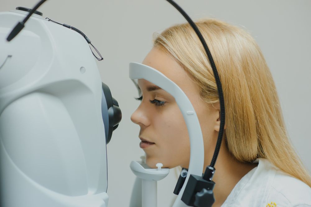
Optical Coherence Tomography
Optical coherence tomography (OCT) is a specialized method for scanning the retina. It creates high-resolution computerized optical images of the retina, similar to viewing slices of the retina under a microscope. The test is painless and does not require bright lights or dilated pupils in many patients. OCT can be helpful for conditions such as macular holes, macular pucker, macular degeneration, and diabetic macular edema.
Other Retinal Diagnostic Tools
In addition to the techniques discussed above, there are many other diagnostic tools retina specialists can use to identify retina and macular conditions, including:
- Farnsworth D15 color test
- Visual evoked potential
- Multifocal electroretinogram
- Full-field electroretinogram
- Electrooculogram
Schedule Your Diagnostic Appointment Today
Advanced diagnostic tools can help diagnose a wide variety of retinal and macular conditions that may otherwise go unnoticed for long periods. Identifying these conditions early can lead to better vision outcomes. At Retinal Consultants Medical Group, we use the most advanced diagnostic tools and equipment to detect retinal issues and develop personalized treatment plans quickly and efficiently. Contact us today for more information or to schedule an appointment with our expert team.